Dataset

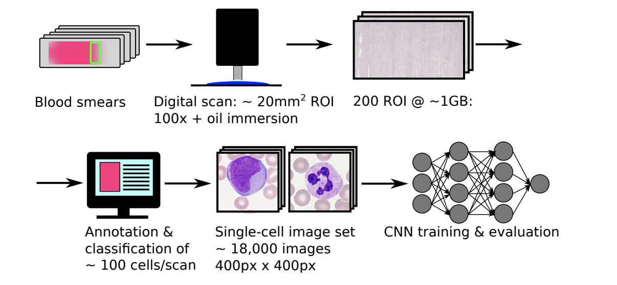
The dataset of single cell white blood cell images we use to train our models comes from a research team in Munich and contains 18,365 images across 15 subtypes of white blood cells (Matek et al., 2019). The data was made available on The Cancer Imaging Archive, and the images were “taken from peripheral blood smears of 100 patients diagnosed with Acute Myeloid Leukemia at Munich University Hospital between 2014 and 2017, as well as 100 patients without signs of hematological malignancy.” The breakdown of these images by class can be seen in table 1 below, and the methodology for the cell imaging can be seen in figure 2 below. It is important to note that myeloblasts are one of the most relevant cells in an AML diagnosis (DiNardo et al., 2016). Additionally, the class imbalances posed some concerns for model training.
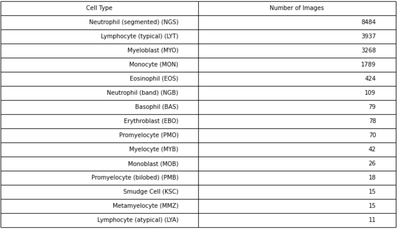
Table 1: Class breakdown of single cell images in Matek et al., dataset. Cell Type includes full name of cell and abbreviation.